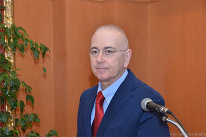
A talk by Prof. Dioguardi at the 2nd Integrative Oncology Conference held in Modena, Italy.
04/03/2018
Watch the video of Prof. Dioguardi’s presentation (in Italian) »
The way in which tumor cells grow and duplicate more efficiently than the normal ones is a very complicate subject from a methodological point of view, and therefore a topic which has not been studied as much as that relating to the attempts to kill them with chemotherapies or other physical and biological tools.
At the very beginning of the chemotherapy (CT) era, in the early Fifties of the XIX century, it was evident that nutrition plays a role both in slowing down the tumor development and in helping the animals to survive to CT. Even if most of the researchers who studies the metabolism of the neoplastic cell consider that anaerobic glycolysis plays a crucial role in the energy production of the tumor (the so-called Warburg effect), very few things are known about the mechanisms regulating the protein and lipid synthesis needed by the cancer cells to divide and spread.

The role of the energy production is essential exactly because the costs for the syntheses are huge, in a dividing cell, but the metabolic advantage of the cancer cell has not been clarified yet. In mammals’ metabolism there is a hierarchy among macronutrients, since the amino acids (AAs) provided by the proteins to the body are the only source of nitrogen, while both AAs and carbohydrates and lipids are sources of carbon, oxygen, and hydrogen. However, AAs are not all the same and proteins contain variable quantities of each AA according to their specific composition.
The most relevant categorization for biological aims is that between essential AAs (EAAs) and non-essential AAs (NEAAs). EAAs are not synthetized by the organism in amounts enough for living, while this happens for NEAAs when enough quantities of EAAs are available, since these latter allow the synthesis of NEAAs by donating part of their molecules. The carbon skeleton of most of the more abundant NEAAs in the proteins of our body, as glutamine, comes from the glucose metabolism in the mitochondrion, that is from transformations happening during the energy production in the Krebs cycle. Interestingly, the bowel is built to destroy the glutamine contained in the foods, it defends us from absorbing glutamine, even if this produces a substance which is potentially dangerous and highly toxic for the brain in case of liver failure, while it helps the absorption of precursors allowing the synthesis where needed in tissues and organs.
Knowing the complicate intra-organs metabolism, based on glutamine, is important because the cancer cells, as well as some cells of our body, particularly those of the immune system, depend and use glutamine as the main source of energy, consuming significant quantities of it to survive and carry on their tasks. Indeed, a lot of therapeutic attempts aimed at blocking the availability of glutamine to the cancer cell are under study. These attempted therapy act on the catabolic mechanisms driven by two specific enzymes, the glutaminases, which have the same substrate (i.e. glutamine) but different outcome products entering in the metabolism.
The growing knowledge on the biological and molecular basis of cancer cells functioning has led to the development of therapies which are very effective when the pharmacologically active dose is reached. However, this dose is often much toxic for organs and tissues, e.g. heart, which have a high metabolic activity, like the tumor. It is thought that 60% of patients do not take a dose able to optimize CT efficiency, and hence to obtain the desired outcomes. Moreover, in these patients it has been observed that CT toxicity is proportional to the integrity of the muscular mass, and the deficiency of peripheral muscle, i.e. sarcopenia, increases the chance of not reaching adequate therapeutic doses. Epidemiological data show that regular physical activity not only reduces the risk of neoplasia and promote longevity, but that it is also associated to a better tolerance to CT drugs and to a reduced number of relapses. Physical activity is the most important factor promoting muscular masses’ integrity, but an adequate diet is a necessary complement for maintaining or promoting the muscular structures. And even if with its specific characteristics, the heart is a muscle. The synthesis of muscle proteins is highly regulated by the intake of AAs, and EAAs (particularly some of them) have a prominent role in triggering this synthesis, when present in adequate quantity. However, protein synthesis is also associated to a very significant energy consumption (in fact, 4 ATPs are required for each AA added to the chain) and to the need to eliminate worn out proteins – and both these conditions are made effective by regular exercise.
There are two main mechanisms driving the elimination of “old” molecules in the cells. Hence, these mechanisms, proteolysis and autophagy, are not valid just for the proteins. They are complex ways in the activation and inactivation processes, highly modulated in the long chain of steps which creates them and activate or inhibit them. Since its recent discover, autophagy has been extensively studied for its high precision of functioning and for the possibility to recycle part of the amino acid component.
Diet has a deep impact on both protein synthesis and proteolysis and autophagy.
My hypothesis is that the neoplastic cell has a peculiar efficiency, and hence an advantage over the normal cell, which is a heritage and conditioning of the environment where the tumor develops and grows. The tumor tends to reduce the energy production in the mitochondrion and privileges the cytoplasmic glycolysis. However, in so doing it significantly reduces the availability of intermediates of the Krebs cycle which can be used to produce NEAAs. Hence, it depends on and consume the NEAAs present in the environment in which it develops, and that are always largely prevalent over the EAAs needed to activate the synthesis. The advantage of using the NEAAs present in the body (i.e. acting as a body parasite) makes it also frail and adapt to that specific environment in which NEAAs are always and invariantly predominant versus those EAAs which are needed to give it the growth rhythm. What happens if the ratio between EAAs and NEAAs is inverted in the environment surrounding the tumor?
Our research has led to discover that in a different environment, with normal cells having maximum adaptability and efficiency, the presence of more EAAs vs. NEAAs inhibits proteolysis, but activate autophagy in tumor cells, particularly in those at high duplication speed, because the cancer protein synthesis needs to face the sudden deficiency of NEAAs – a relative deficiency compared to the abundance of EAAs. This environmental change, which is extremely beneficial for the normal cell, highlights a selective weakness of the tumor cell – its inability to adapt to environmental changes. Therefore, to preserve a quantity of NEAAs adequate to duplicate all the proteins, and sustained by the abundance of EAAs, autophagy triggers a destructive mechanism for the cell in the precise moment in which it is duplicating, hence activating a cellular suicide method, i.e. apoptosis, as we have described in more detail in a recent paper.
We had previously proved that supplementation with EAAs has a protective effect against the damage caused by CT drugs on both heart and kidney. Subsequently we showed that AAs supplementation increases the effects of CT on the cancer cell. Now we have the in vitro demonstration of the specific lethal effect of the change of EAAs/NEAAs ratio on the neoplastic cell, and vice versa of its benefit for the normal cell.
Is there a clinical future for the use of important modifications of the diet quality? Potentially yes, but at present it is difficult to foresee how far the discovery of such specific metabolic weakness of the neoplastic cell can lead. It is an interpretation key of cancer metabolism which could have extraordinary developments – it opens a completely new direction in the approach to therapy and fight against the tumor. However, our work also highlights the error of having made so slow, difficult and expensive the research on animal models here in Italy, obliging the substantial developments to be transferred outside the country.
Prof. F.S. Dioguardi
Gastroenterologist, Associate Professor of Internal Medicine, Department of Clinical Sciences and Community Health, University of Milan, Italy

